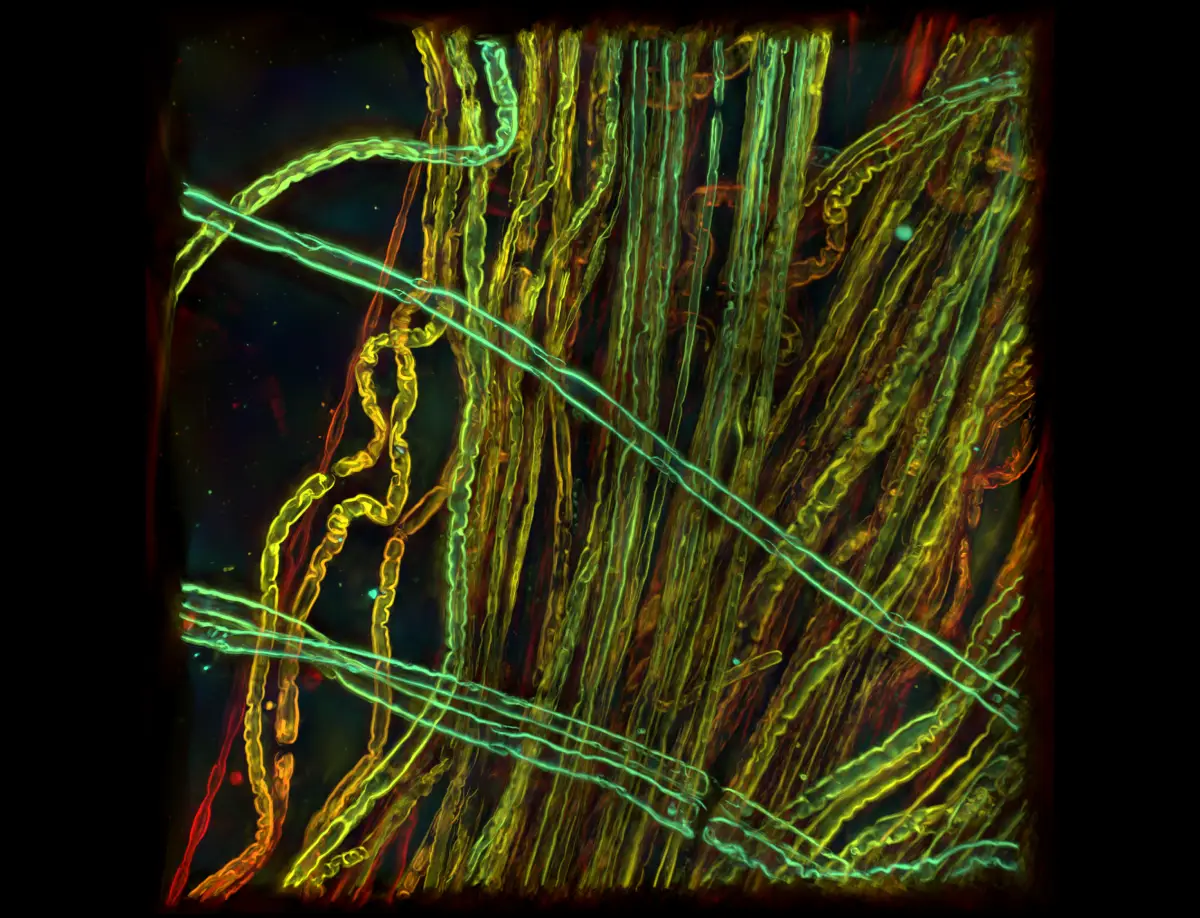
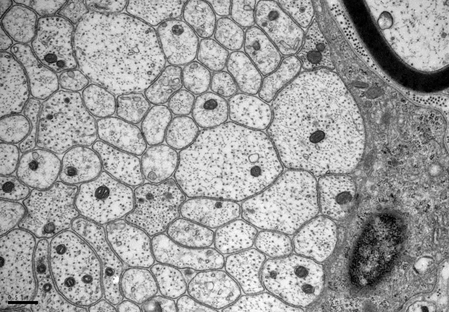
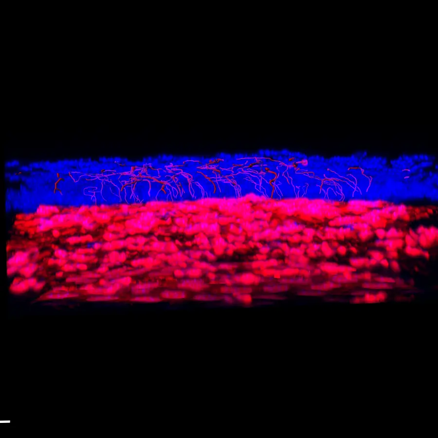
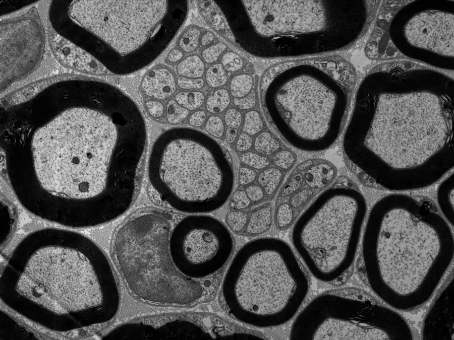
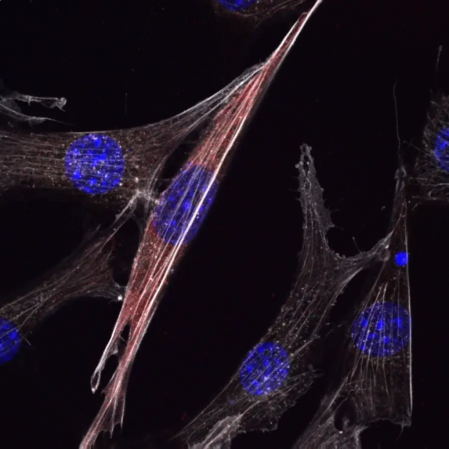
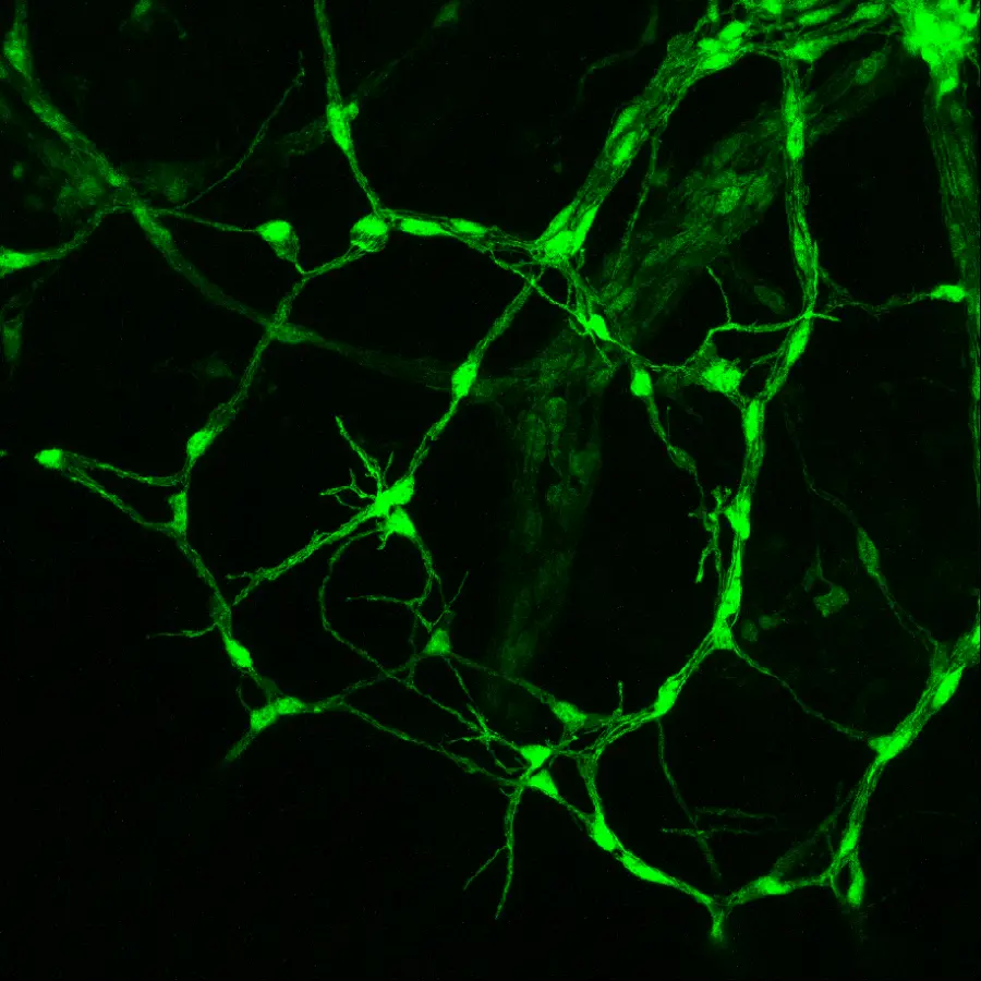
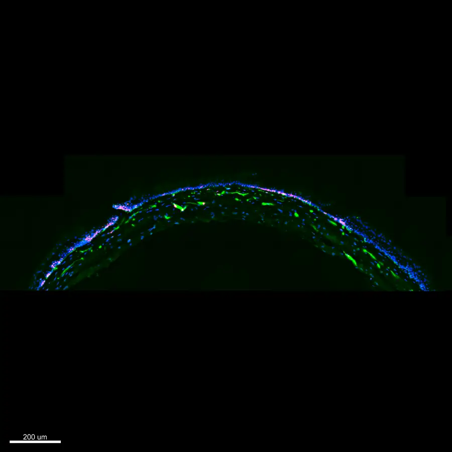
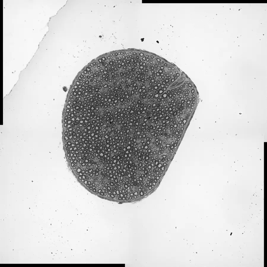
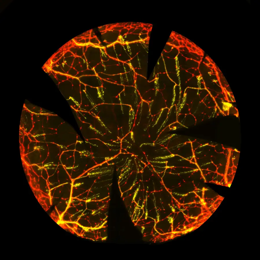
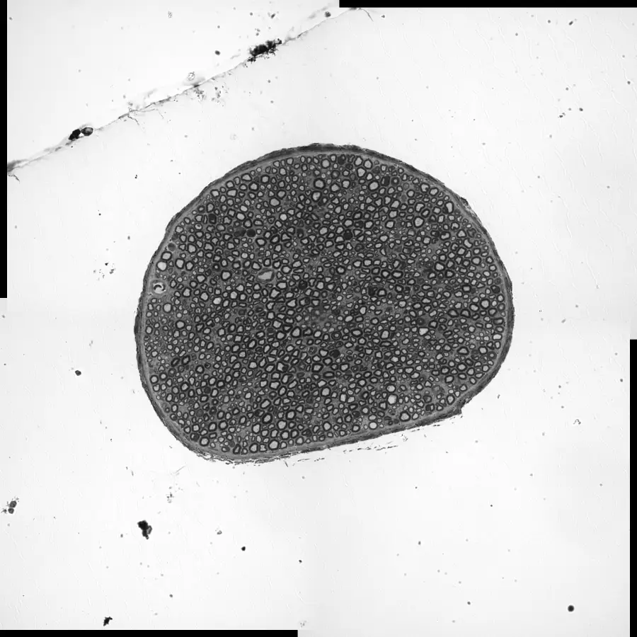
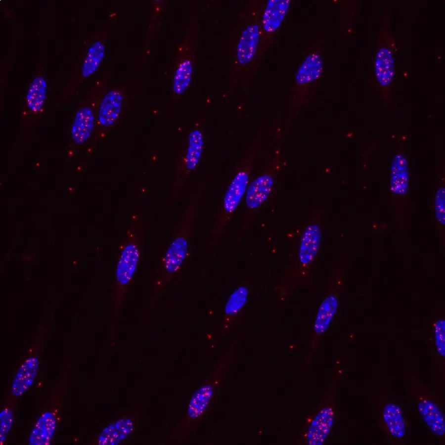
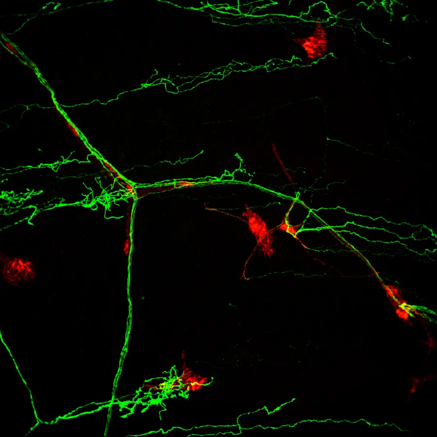
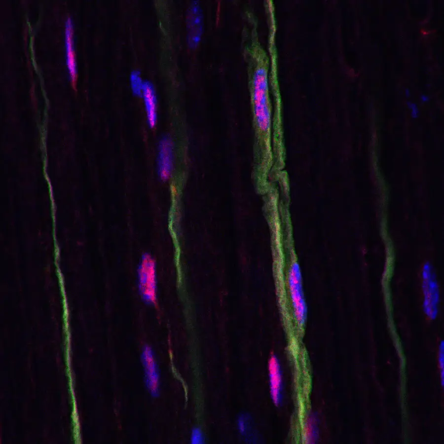
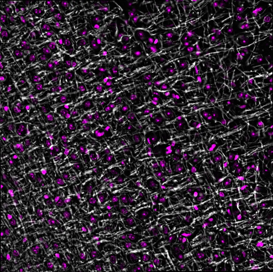
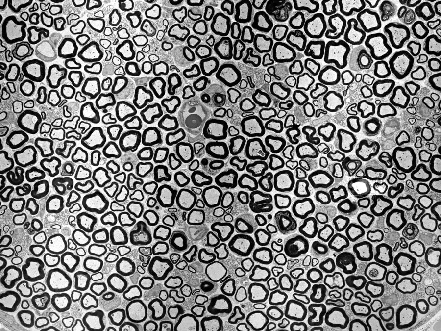
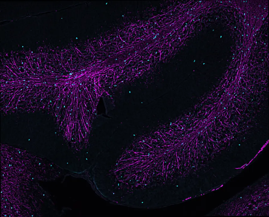
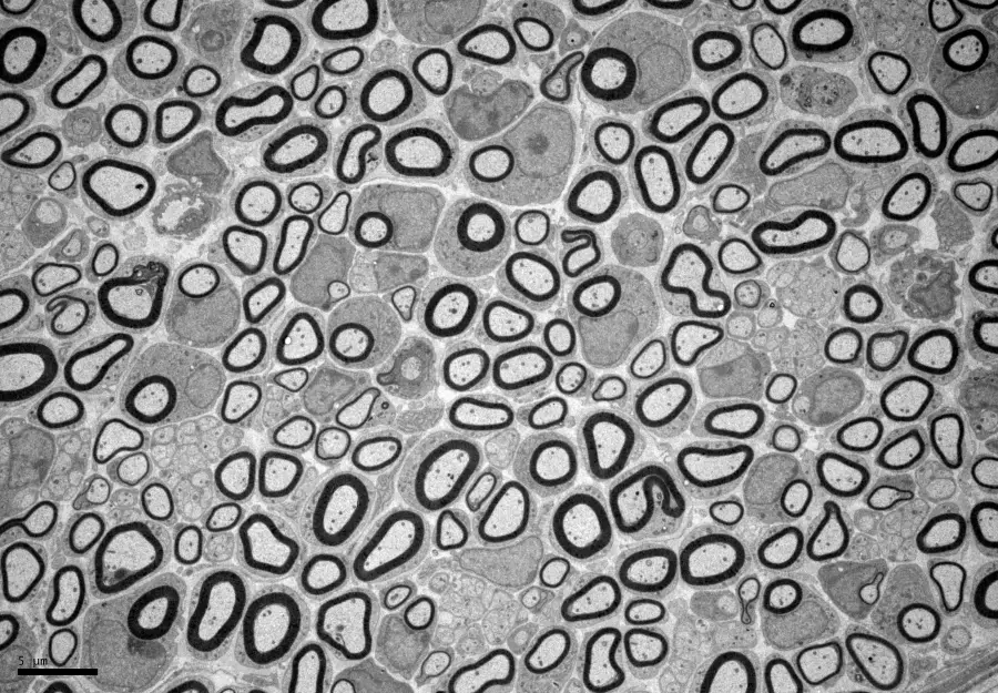
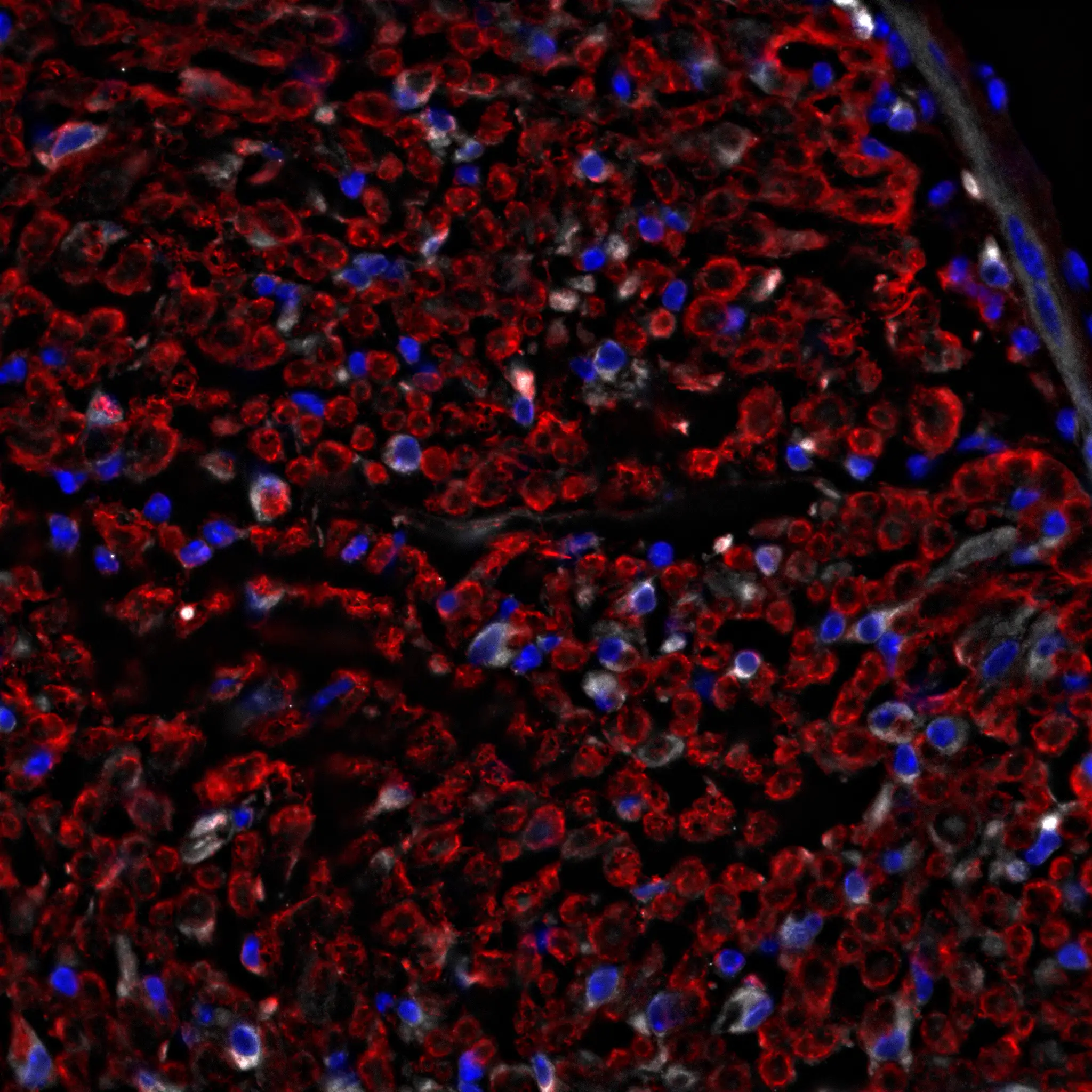
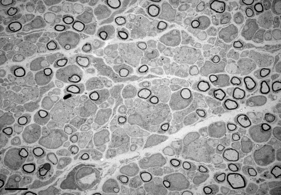
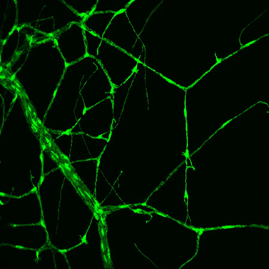
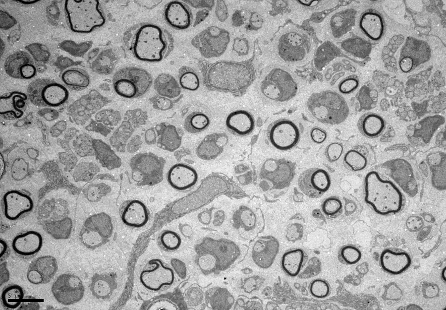
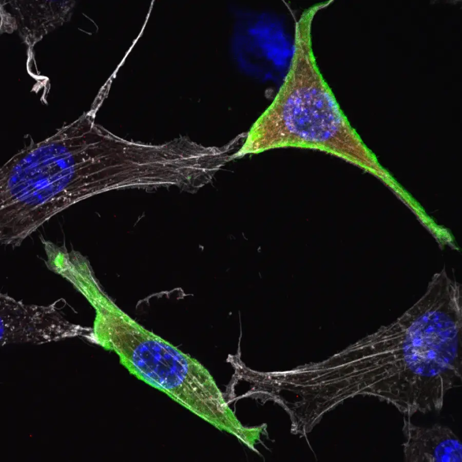
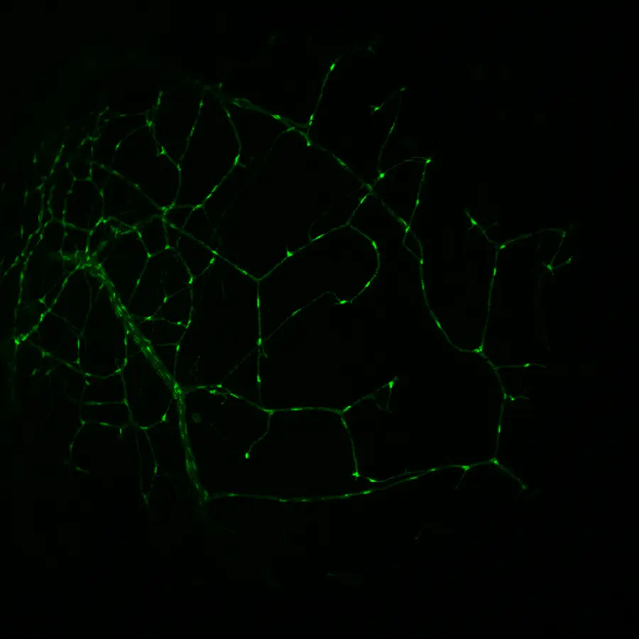
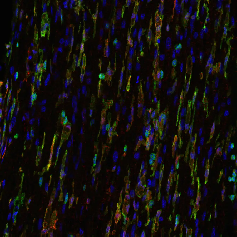
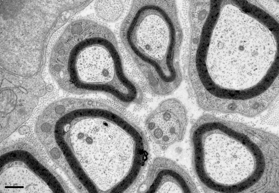
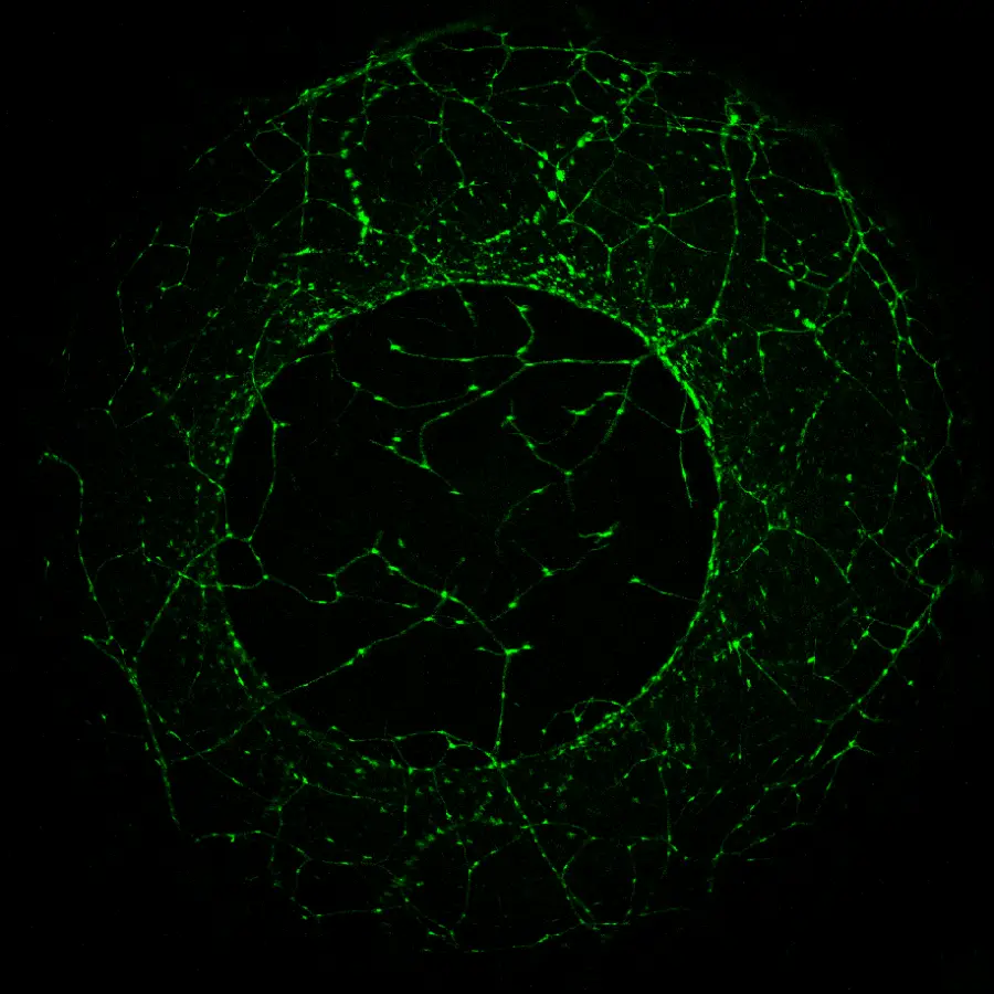
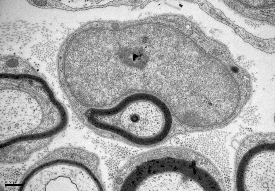
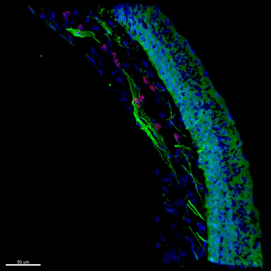
In the Myelin Alicante Labs gallery, you can explore high-resolution images obtained through advanced microscopy techniques. The brightfield microscopy images reveal detailed structures of the peripheral and central nervous systems, while fluorescence images highlight specific glial cells, such as myelinating glia. Confocal microscopy allows for 3D visualization of nervous tissue sections, offering deeper insights into cell interactions.
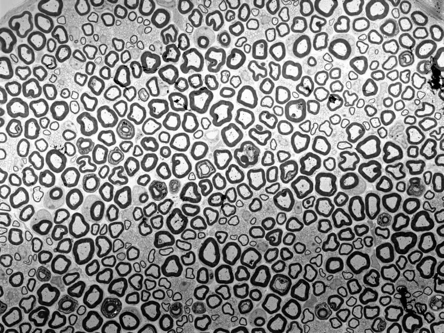
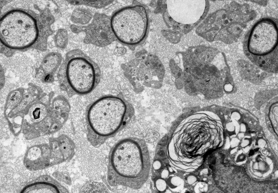
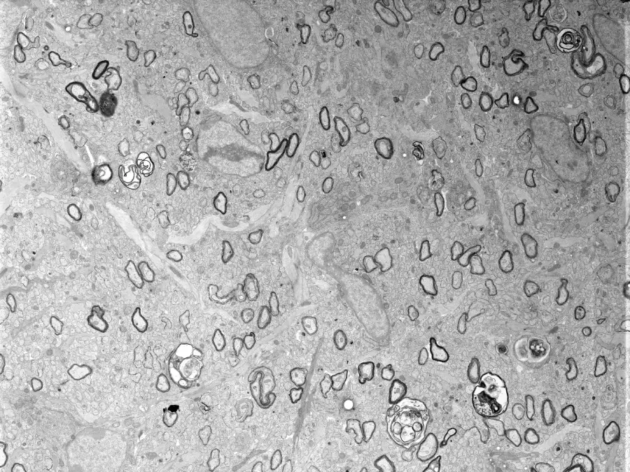
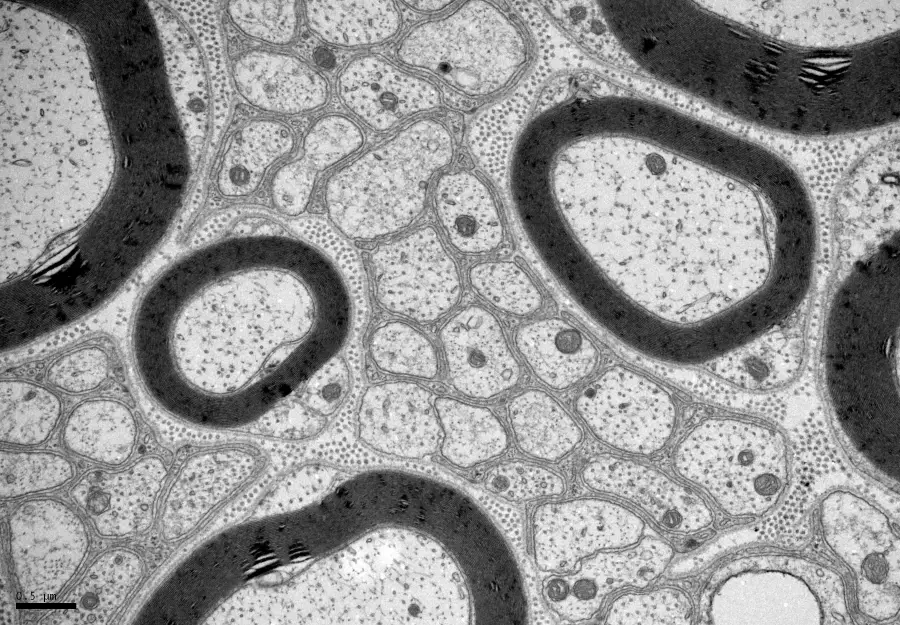
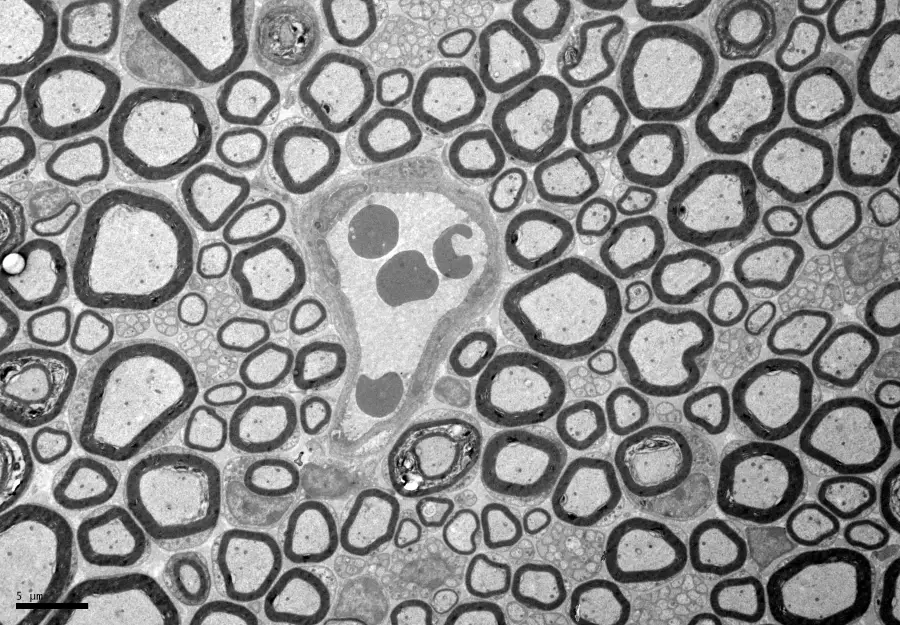
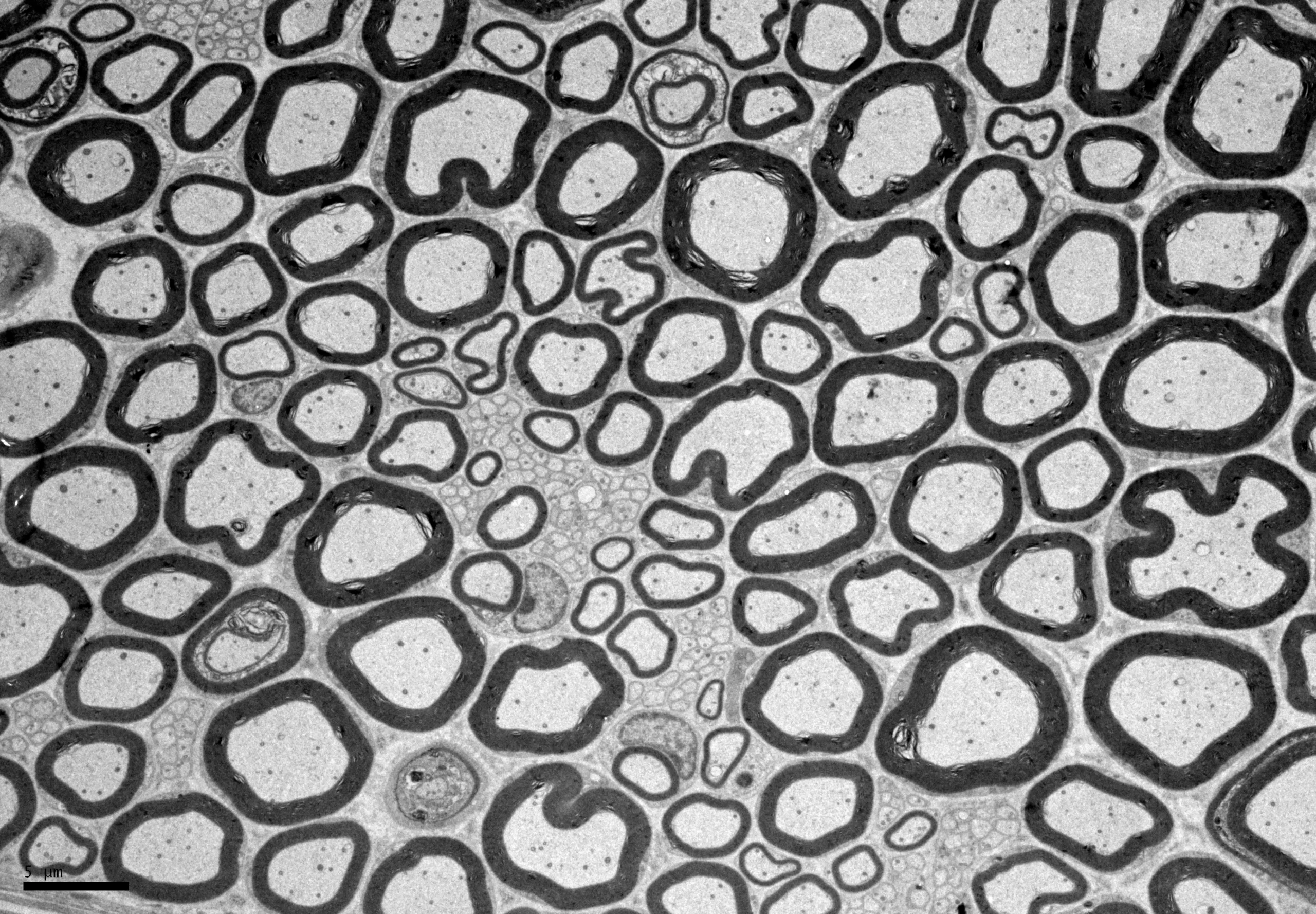
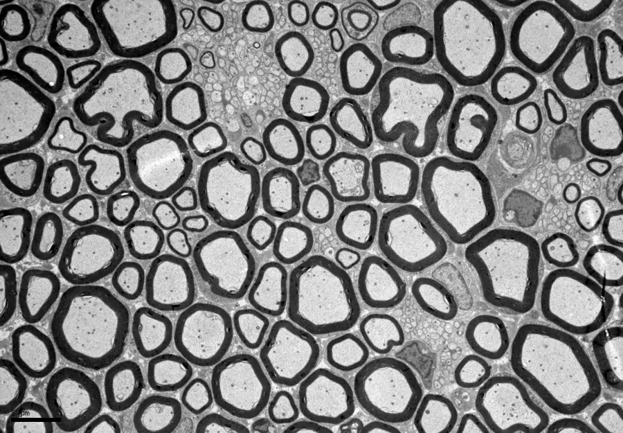
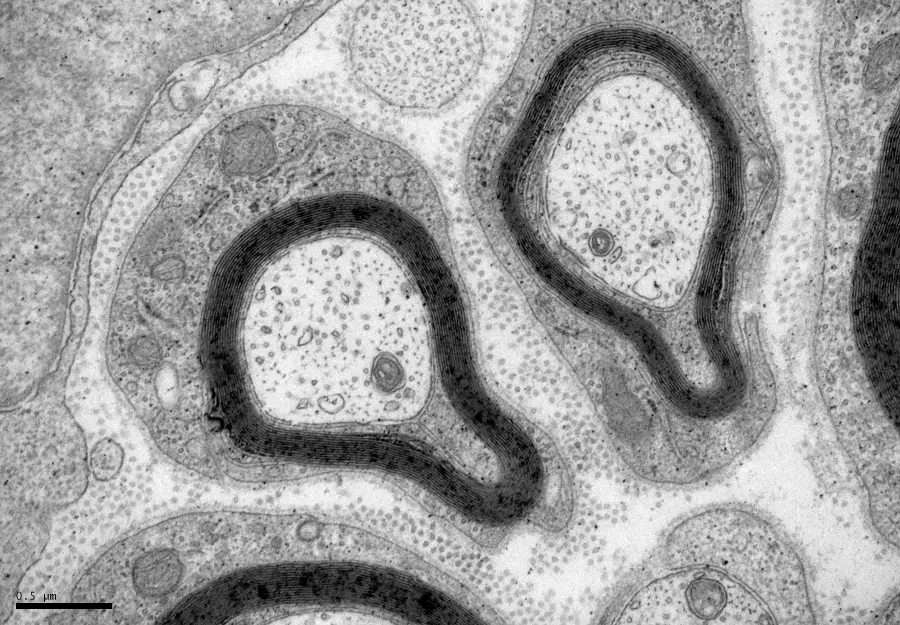
Additionally, electron microscopy images uncover the ultrastructure of axons and myelin, providing details not visible through other techniques. The samples also include cornea tissue, and the study focuses on key glial cells like Schwann cells, oligodendrocyte precursor cells (OPCs), and oligodendrocytes.