The traditional view of the heart as a simple mechanical pump is officially obsolete. Understanding the active role of cardiac glia marks the beginning of a new era in neurocardiology, paving the way for precise biomarkers and novel anti-arrhythmic therapies.
Moving Beyond Neurons in the Heart
For decades, cardiology held an exciting truth: the heart possesses its own «little brain»—an autonomous neural network capable of adjusting the heartbeat millisecond by millisecond. However, scientific attention was focused almost exclusively on neurons. Everything changed when recent discoveries in neurocardiology revealed that the true coordinators of the intracardiac nervous system do not work alone.
Enter cardiac glial cells. Historically considered mere structural «glue»—a term derived from the Greek word glia—we now know that these cells actively regulate heart rate, guide nerve development, and respond dynamically to cardiovascular disease. In this science outreach guide, we explore how cardiac glia is set to revolutionize modern medicine.
What is Cardiac Glia and Why is it Transforming Neurocardiology?
The cardiac autonomic nervous system allows the heart to respond instantly to stress or relaxation through ganglionated plexuses distributed across epicardial fat tissue.
The decisive scientific milestone came from the Human Heart Cell Atlas. Using single-cell RNA sequencing, researchers re-analyzed a cellular cluster previously classified as neuronal, discovering that it lacked neuronal markers and instead expressed pan-glial genes. This discovery proved that the glial population in the heart is extraordinarily abundant and diverse.
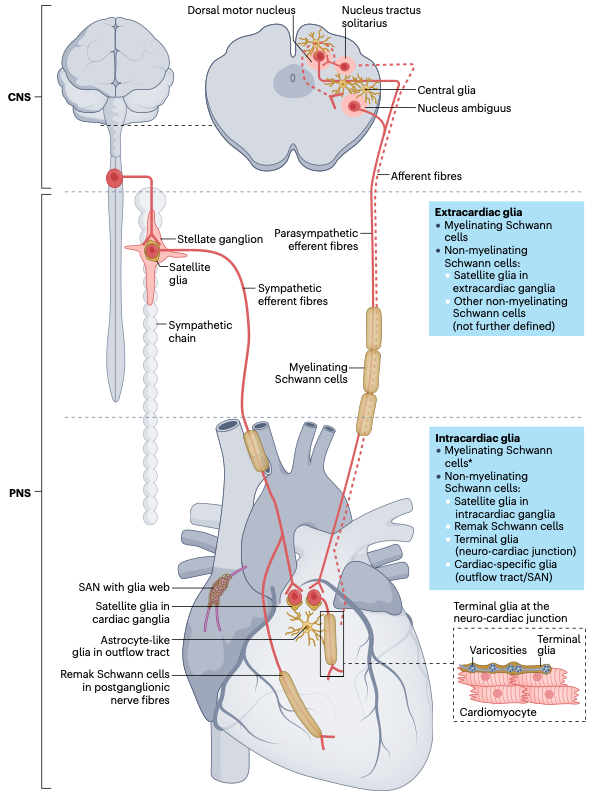
2. The 5 Specialized Subtypes of Cardiac Glial Cells
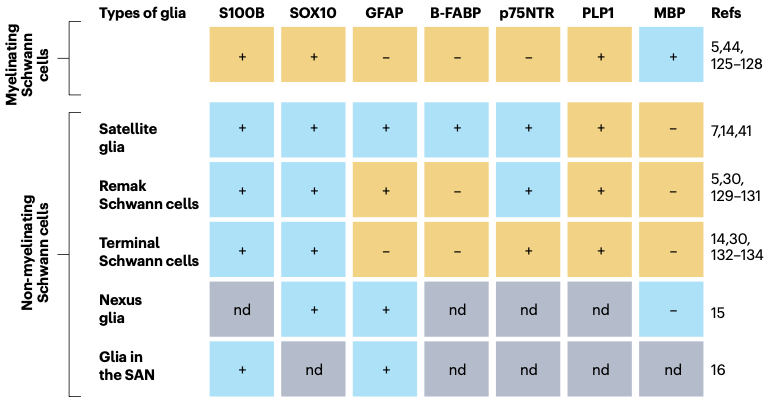
Cardiac glia constitutes a heterogeneous population where each subtype performs a unique functional role:
A. Satellite Glial Cells
Located within intracardiac ganglia, they closely envelop the cell bodies of cholinergic neurons. They supply crucial nutrients, regulate the ionic microenvironment, and participate in parasympathetic innervation development. In human heart immunohistological stainings (Figure 1c of the study), they are identified by the S100B protein (green fluorescence) surrounding neurons expressing choline acetyltransferase (ChAT, in red).
B. Remak Schwann Cells
These non-myelinating cells coat small-caliber unmyelinated C-fibers. Expressing periostin, they are essential for guiding the guidance and fasciculation of sympathetic nerves during embryonic development.
C. Terminal (Perisynaptic) Glia
Positioned at the neurocardiac junction, terminal glia operates within a «tripartite synapse,» releasing neurotrophic factors (BDNF and GDNF) that stimulate Neuregulin 1 to maintain cell plasticity and synaptic stability.
D. Nexus Glia (Astrocyte-Like)
Found in the cardiac outflow tract, these cells exhibit star-like morphology and express GFAP. They modulate the heart’s response to autonomic stimulation; their absence leads to severe arrhythmias such as ventricular tachycardia.
E. Interstitial Glial Network of the Sinoatrial Node (SAN)
Intertwined directly with pacemaker cells, this network generates intracellular calcium oscillations that help initiate and stabilize the rate of every heartbeat.
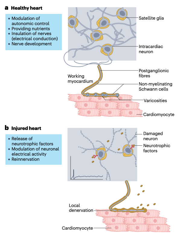
3. Pathophysiology: When Glia Drives Arrhythmias
In conditions such as myocardial infarction, atrial fibrillation, or diabetes, glial cells undergo a pathological remodeling process called reactive astrogliosis.
During reactive gliosis, glia becomes hypertrophic and increases GFAP expression. Although initially an acute protective response, persistent activation triggers pathological cascades:
- Sympathetic Hyperactivity: Excessive glial activation in the stellate ganglion induces massive norepinephrine release, heightening the risk of ventricular arrhythmias.
- Arrhythmogenic Axonal Sprouting: Following injury or denervation, glia releases neurotrophic factors that stimulate chaotic nerve sprouting, creating an arrhythmogenic substrate.
4. Clinical Applications: S100B and Novel Therapies
The study of cardiac glia directly translates into actionable clinical tools:
- S100B as a Diagnostic Biomarker: In catheter ablation procedures for atrial fibrillation, neurocardiac injury causes immediate release of S100B into the bloodstream, serving as a precise indicator of autonomic damage.
- Neuregulin 1 in Heart Failure: Clinical trials have demonstrated that Neuregulin 1 administration improves left ventricular function and reduces pathological sympathetic tone.
- Targeted Glial Modulation: Developing drugs that specifically target glial cells represents a promising frontier for stabilizing cardiac rhythm without systemic side effects.
Reference: Scherschel, K., et al. (2025). Glia of the heart’s nervous system. Nature Reviews Neuroscience.