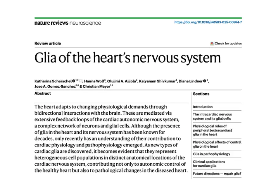
Recent advances in neurocardiology highlight that glial cells within the cardiac nervous system are not merely passive structural elements, but active regulators of cardiac function, development, and pathology.
Single-cell transcriptomic analyses and functional studies demonstrate that cardiac glia form a highly heterogeneous population—comprising satellite glia, Remak Schwann cells, terminal glia, astrocyte-like nexus glia, and sinoatrial node glial networks—that interact directly with autonomic neurons to modulate heart rate and signal transmission.
Furthermore, under pathophysiological conditions like atrial fibrillation and myocardial infarction, these cells undergo reactive remodeling, release neurotrophic biomarkers such as S100B, and actively participate in pro-arrhythmic sympathetic signaling, opening promising avenues for novel diagnostic and therapeutic strategies.
Discovery and Cellular Diversity of Glia in the Cardiac Nervous System
Cardiac glial cells represent a highly heterogeneous population that decisively expands our understanding of the «little brain of the heart.»
Although neurocardiology historically focused on autonomic neurons, recent single-cell transcriptomic analyses from the Human Heart Cell Atlas have demonstrated that cell clusters previously classified as neuronal actually correspond to glial cells.
Within the intracardiac nervous system—located primarily in the ganglionated plexuses of epicardial fat—multiple specialized subtypes coexist: satellite glial cells wrapping cholinergic neuronal somas, Remak Schwann cells protecting unmyelinated fibers, and terminal glial cells positioned at neurocardiac junctions.
In addition, unique subpopulations have been identified, such as nexus glia (exhibiting astrocyte-like morphology in the outflow tract) and an interstitial glial network in the sinoatrial node, confirming that cardiac glia possess an anatomical and molecular organization tailored to the specific needs of each heart region.
Physiological Mechanisms and Neuroglial Modulation of Cardiac Function
Far beyond providing trophic and structural support, cardiac glia actively participate in the development of innervation and the dynamic regulation of cardiac rhythm.
Satellite glial cells supply nutrients to intracardiac neurons and facilitate the guidance and fasciculation of autonomic nerves from embryonic stages. Meanwhile, the glial network in the sinoatrial node is closely intertwined with pacemaker cells, participating in intracellular calcium oscillations that help initiate and stabilize heart rate.
Likewise, nexus glial cells interact directly with sympathetic and parasympathetic innervation in the outflow tract, acting as essential modulators of the chronotropic response to autonomic stimuli.
Pathophysiological Relevance, Biomarkers, and Clinical Perspectives in Neurocardiology
Under pathological conditions such as myocardial infarction, atrial fibrillation, and heart failure, glia undergo reactive remodeling that directly impacts the arrhythmogenic substrate.
Following neurocardiac injury caused by catheter ablation or local denervation, intracardiac glia act as first responders by releasing neurotrophic factors such as the S100B protein, which stimulates axonal growth and alters neuronal electrophysiological properties.
Clinically, plasma levels of S100B have emerged as a key biomarker for assessing the extent of neurocardiac injury during atrial fibrillation ablation. Furthermore, because glial hyperactivity in stellate and intracardiac ganglia contributes to heightened pathological sympathetic tone and promotes ventricular arrhythmias, selective modulation of cardiac glia represents a promising therapeutic target for restoring autonomic control of the heart.
Explore the full paper to unlock the latest insights into the ‘little brain’ of the heart.